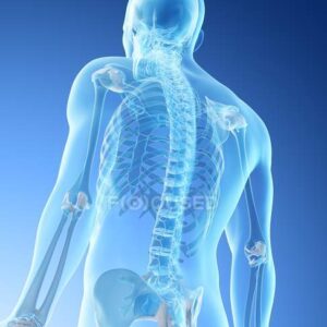
The skeletal system comprises Periosteum, cartilage and bones( osseous tissue).
The bones support soft tissue, protect internal organs and act as a rigid lever for movement.
Bones also act as an important source of nutrients and blood constituents.
The skull, vertebral column, sternum and ribs are called as the axial skeleton.
The bones of the upper and lower limbs along with their respective girdles are called appendicular skeleton.
Axial Skeleton
Skull
Vertebral Column
Ribs
Sternum
Hyoid Bones
Appendicular Skeleton
Upper Limbs
Scapula
Clavicle
Humerus
Radius & Ulna
Carpals & Metacarpals
Phallanges
LowerLimbs
Pelvis
Femur
Humerus
Tibia & Fibula
Tarsal & Metatarsal
Phallanges
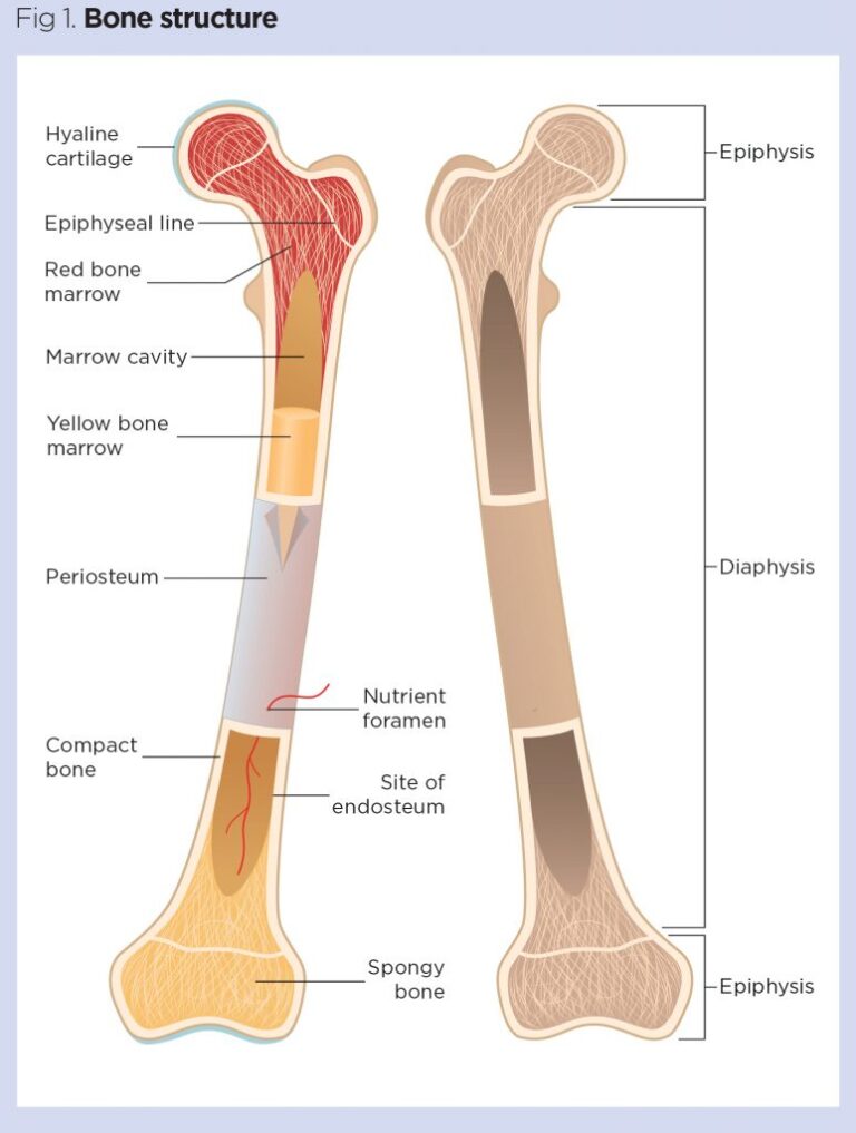
Bone Structure:-
The main portion of a bone or shaft is called the “diaphysis”. The medullary cavity, or marrow cavity, is the space inside the diaphysis.
The marrow cavity is covered by a layer of endosteum, which contains cells necessary for bone development. The ends of the bone are called the “epiphyses”.
The region of mature bone where the diaphysis joins each epiphysis is called the “metaphysis.”
The epiphyses are covered by articular cartilage. Cartilage is a semirigid form of connective tissue that helps to reduce friction and absorbs some of the shock in synovial joints.
The periosteum is a membrane covering the surface of bones, except at the articular surfaces. The periosteum is composed of two layers, an outer fibrous layer and an inner highly vascular layer that contains cells for the creation of new bones. The periosteum is the point where ligaments and tendons get attached and is critical for bone growth, repair, and nutrition.
Bone Facts:-
1. Number of Bones: 206
2. Number of Bones in Newborn : 306
3. Number of vertebrae in the neck : 7
4. Number of Ribs : 24 ( 12 pairs)
5. Number of vertebrae : 33
6. Number of bones in face : 14
7. Number of bones in skull : 22
8. Number of bones in Chest : 25
9. Number of bones in arm :6
10. Longest bone : femur
11. Smallest Bone : Stapes middle ear
Bone Classified:-
Bones can be classified into 4 types based on their shapes:-
Long Bones ( Radius, Ulna, Tibia, Femur and Humerus)
Flat Bones (Sternum, Pelvis, Ribs, scapula)
Short Bone (Carpals and tarsals)
Irregular Bone ( Vertebrae, sacrum, coccyx)
The human bone is composed of the following
Periosteum
Compact Tissue
Spongy Tissue
Bone Marrow
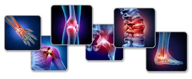
Articular System: Joints
Joints are classified as synarthrodial, amphiarthrodial, or diarthrodial (synovial)
Joints are classified as synarthrodial, amphiarthrodial, or diarthrodial (synovial)
Synarthrodial joints -Do not move. These are fibrous joints. ( Example: sutures of the skull.)
Fibrous joints contain fibrous connective tissue and cannot move; fibrous joints include sutures, syndesmoses, and gomphoses.
Amphiarthrodial joints – move slightly and are held together by ligaments (e.g., inferior tibiofibular joint)
Cartilaginous joints contain cartilage and allow very little movement
Synarthrodial and amphiarthrodial joints do not contain an articular cavity, synovial membrane, or synovial fluid.
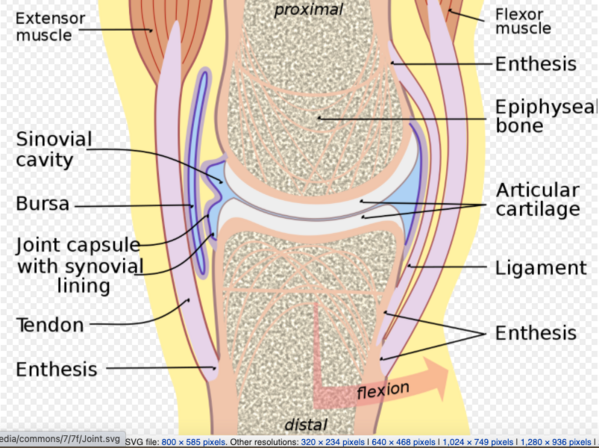
Synovial Joint:-
Synovial joints are the most common type of joint in the body . These joints are diarthrodial, meaning they are freely mobile.
A characteristic for a synovial joint that is not seen at fibrous or cartilaginous joints is the presence of a joint cavity. The joint cavity contains synovial fluid, secreted by the synovial membrane (synovium), hich lines the articular capsule. The articulating surfaces of the bones contact each other in this fluid-filled space. Hyaline cartilage forms the articular cartilage, covering the entire articulating surface of each bone.
A few synovial joints of the body have a fibrocartilage structure located between the articulating bones. This is called an articular disc, which is generally small and oval-shaped, or a meniscus, which is larger and C-shaped. Synovial joints are further classified based on the type of movements they allow.
There classifications are: hinge (elbow), saddle (carpometacarpal joint), planar (acromioclavicular joint), pivot (atlantoaxial joint), condyloid (metacarpophalangeal joint), and ball and socket (hip joint)
The bones in a synovial joint are connected by ligaments.
Ligaments are a type of tough, fibrous and elastic connective tissue. Ligaments appear as crisscross bands that attach bone to bone and help stabilize joints.
They aprevent dislocation by restricting actions outside the normal range of motion. They also absorb shock because of their elasticity, which protects the joint from unwanted external injury.
They help maintain posture and movement.
The movement at a synovial joint is caused by the muscles attached across the joint. Muscles are attached to bone by tendons. Tendons are very strong, inelastic connective tissues that allow a muscle to pull on a bone to move it. Tendons are found throughout the body, from the head down to the feet. The Achilles tendon is the largest tendon in the body. It attaches the calf muscle to the heel bone.
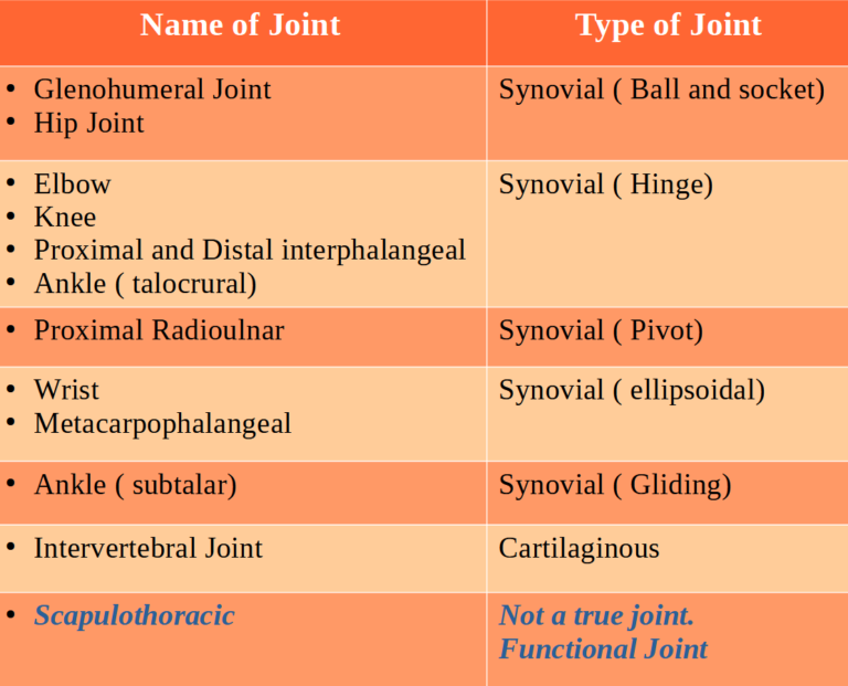
Names of Joints and their types:-
Joints Movements:-
A combination of rolling, sliding, and spinning of the joint surfaces is considered as joint movement.
“Open chain” movements occur with the movement of the distal segment of a joint. Example: leg extension exercise on a machine causes open chain movement for the knee joint.
“Closed chain” movements occur when the distal segment of the joint is fixed in space. Example: Action of the knee joint in standing barbell squats movement.
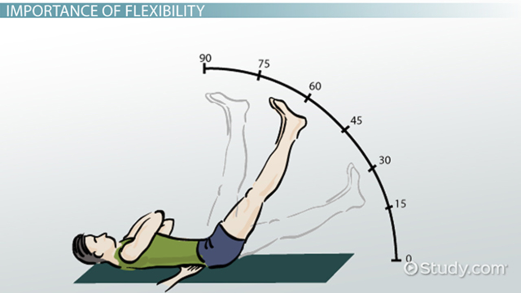
Range of Motion:-
Range of motion is the capability of a joint to go through its complete degree
“Open chain” movements occur with the movement of the distal segment of a joint. Example: leg extension exercise on a machine causes open chain movement for the knee joint.
“Closed chain” movements occur when the distal segment of the joint is fixed in space. Example: Action of the knee joint in standing barbell squats movement.
Passive range of motion can be defined as the range of motion that is achieved when an external force causes movement of a joint and is usually the maximum range of motion that a joint can move.
Active range of motion is the range of motion that can be achieved by voluntary movement from contraction of the associated skeletal muscles. For example, the active range of motion to allow the elbow to bend requires the biceps to contract while the triceps muscle relaxes.