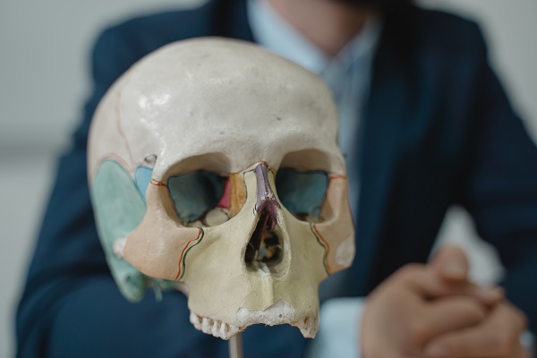
The Skull and the Bones of the Head:-
The Head comprises of the skull , face and brain.
The main function of the skull is the protection of the most important organ in the human body, the brain. Also, the skull provides support for all of the facial structures.
- The skull is composed of two parts: the cranium and the mandible.
- The skull has 22 bones out of which 8 bones form the Cranium and 14 bones form the face.
Components & Features
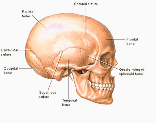
- The bones of the skull are joined together by ossified joints called sutures.
- The skull is divided into the braincase (neurocranium) and the facial skeleton(viscerocranium).
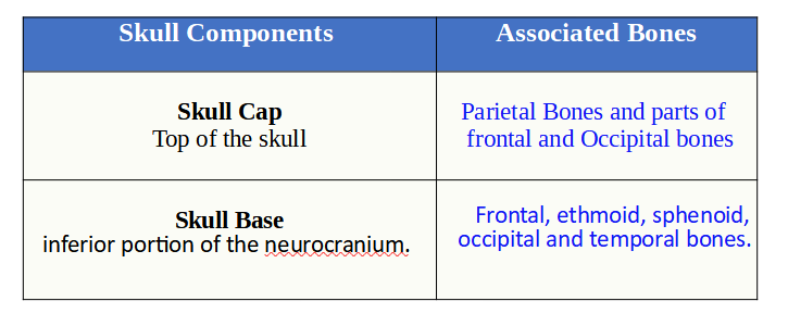
- The brain-case consists of the skullcap (calvarium) and the skull base. The brain is enclosed by the neurocranium. Most foramina in which nerves and blood vessels pass through are located at the base of the skull.
The largest opening in the skull is the foramen magnum. Here the brainstem
( MEDULA OBLONGATA) leaves the skull and becomes the spinal cord.
The bones in the skull can be divided into the
Cranial bones:- which form your cranium ( 8 nos)
Facial bones:- which make up the face. (14 nos)
The 8 bones in the cranium forms a “Vault” which encloses and protects the brain.
8 bones in the cranium
- Frontal Bones
- 2 nos of Parietal Bones
- Occipital Bones
- 2 nos of Temporal Bones
- Sphenoid Bones
- Ethmoid Bones.
14 Facial Bones
- Vomer Bones
- 2 nos of Nasal Conchae
- 2 Nasal Bones
- 2 nos of Maxillae
- Mandible
- 2 nos of Palatine
- 2 nos of Zygomatic Bones
- 2 nos of Lacrimal Bones
Parts of the Skull Base
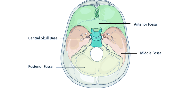
The skull base is the inferior part of the neurocranium. It can be divided into the anterior, middle and posterior fossae.
The Anterior Cranial Fossa
It is a depression in the floor of the cranial base which houses the frontal lobes of the brain. The anterior cranial fossa consists of three bones: the frontal bone, ethmoid bone and sphenoid bone.
The Middle Cranial Fossa
It lies slightly deeper than the anterior cranial fossa. It is “butterfly shaped”, with a middle part accommodating the pituitary gland and two lateral parts accommodating the temporal lobes of the brain. In this part, the greater and lesser wings of the sphenoid bone contains the trochlear nerve, abducens nerve, oculomotor nerve and ophthalmic nerve.
The Posterior Cranial Fossa
It is the most posterior part of the skull base containing the brainstem and cerebellum. The posterior cranial fossa is comprised of three bones: the occipital bone and the two temporal bones.
The frontal bone is located superiorly in the skull and underlies the forehead; above the orbital cavities, the nasal bridge, and the frontal process of the zygomatic bone. It protects the frontal lobes of the brain.
The posterior part of the skull is formed by the parietal bone . It forms a part of the side and top of the head. In front each parietal bone joins with the frontal bone. In the back, they connect with the occipital bone and below with the temporal and sphenoid bones.
The posterior aspect of the skull is formed by the occipital bone. The occipital bone is a trapezoid-shaped bone at the lower-back of the cranium. It supports the back part of the brain. There is a large oval opening in the occipital bone that is called the foramen magnum. The spinal cord passes through this opening. The occipital bone is the only cranial bone to connect to the cervical spine.
The occipital bone connects with the first vertebra,called the atlas and forms the atlantooccipital joint. This joint helps in the movement of the neck.
The temporal bones are paired bones that make up the sides and base of the skull (cranium). Each temporal bone is composed of five parts: the squama, the petrous, mastoid, and tympanic parts, as well as the styloid process . Forming the rear part of the temporal bone, the outer surface of the mastoid part attaches to muscles that regulate the motion of eyebrows and the ear (superior auricular muscle). The temporal bone attaches to the joint of the jaw bone and is fused with other bones of the skull, including the occipital bone on the lower rear side, the parietal bone above that, the sphenoid bone on its front side.
This bone surrounds the middle and inner portions of the ear. Its lower portion connects with the mandible or jawbone and allows the mouth to open and close.
The sphenoid bone lies at the middle base of the skull, of the skull. It is “butterfly shaped”, with a middle part having a cup like depression for accommodating the pituitary gland.
The ethmoid bone is a cube-shaped bone, very light and sponge-like in textured and located in the center of the skull between the eyes. It helps to form the walls of the eye socket, or orbital cavity, as well as the roof, sides, and interior of the nasal cavity.
It helps to support a variety of everyday activities. The cribriform plate has sieve-like holes that allow the passage of the olfactory nerves in the nose so that we can smell and taste things. The nasal conchae that the ethmoid forms allow air to circulate and become humidified as it travels from the nose on the way into the lungs.
Sutures of the Skull
There are several major bones that are connected together to form the skull. These bony plates cover the brain and are held together by fibrous material called sutures.The major bones that compose the skull of a newborn include the following:
2 frontal bones
2 parietal bones
1 occipital bone
Sutures allow the bones to move during the birth process. They behave as expansion joints and allows the bone to enlarge evenly as the brain grows and the skull expands.
Sutures of a New Born's Skull
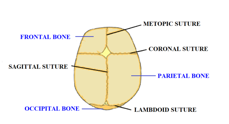
The most important sutures in the human skull are:
- The Metopic suture:- This extends from the top of the head down till the middle of the forehead,.2 frontal bone plates meet at the metopic suture. (variably present in adults )
- The Coronal suture -between the frontal and parietal bones
- The Sgittal suture – between the two parietal bones
- The Lambdoidal suture– horizontally situated between the occipital bone and both parietal bones
Bones of the Face:-
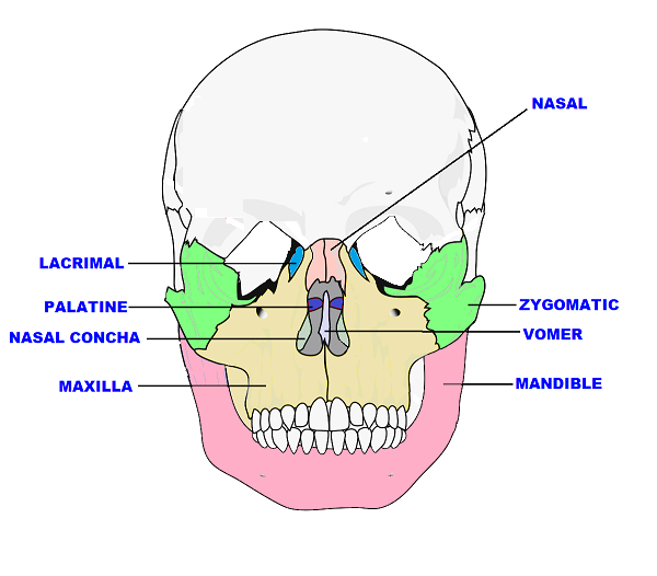
- Nasal – Paired bones forming the structure of the nose.
- Lacrimal – Paired bones that form the wall of the orbit.
- Zygomatic – Paired facial bones that form the cheeks.
- Palatine -Paired, L-shaped bone located posterior to the nasal cavity
- Maxilla – Central, paired bone of the viscerocranium. Left and right maxilla fuse to forms the upper jaw.
- Mandible – Bone that forms the lower jaw.
- Vomer– Single bone of the viscerocranium situated in the midsagittal line. It is a flat plate of bone situated vertically in the nasal cavity.
- Nasal Concha– bony plate located on the lateral wall of the nasal cavity. Its function is the majority of airflow direction, humidification, heating and filtering of air inhaled through the nose.